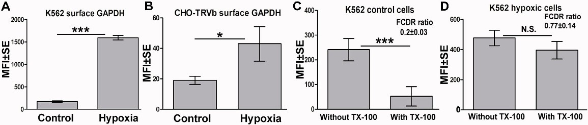
Fig. 2. Membrane recruitment of GAPDH in early stages of hypoxia. (A&B) Cell surface GAPDH exposure is significantly increased after 24 hrs hypoxia treatment in both K562, p<0.0001 (A) and CHO-TRVb, p <0.05 (B) cells. In both cases n=104. (C&D) GAPDH expressed on the plasma membrane of hypoxia treated cells is preferentially recruited to the detergent resistant membrane domain as compared to GAPDH resident on the surface of control normoxic cells. In control cells a significant decrease in surface GAPDH upon extraction with Triton X-100 was observed (p<0.0001, n=104) indicating localization of GAPDH in non DRM portion of membrane. No such loss of GAPDH signal is observed in hypoxia treated cells suggesting migration of GAPDH molecules into the DRM region of membrane (p>0.05, n=104). Representative results are shown and the entire experiment was repeated three times. In each case the FCDR ratios, were calculated using mean flowcytometry based fluorescence intensity measurements as described in materials and methods. The FCDR demonstrated significant increase upon exposure to hypoxia as compared to control cells, p<0.05, n=3.